Tuesday, August 11, 2015

Featured Diagnosis: Dodder (Cuscuta spp.)

A landscape with yellow mats of a parasitic plant covering the plants and ground
Large mats of dodder parasitizing weeds on a disturbed
road side site. (Photo J. French NMSU-PDC) 
Environmental conditions across the state are perfect for the parasitic weed, dodder, to germinate and grow. This parasite does not have leaves and cannot produce chlorophyll. As such, it grows on other plants, using them for water, nutrients, and carbohydrates. It is common along road sides, but can also grow on crop and landscape plants. In the spring, dodder seeds germinate near the soil surface and send up slender, thread-like twining stems varying in color from pale green to yellow or orange and without any cotyledons (seed leaves). The slender, leafless, thread-like stem sways or rotates slowly until it touches the stem or leaf of another plant and begins to wind around it. On a host plant, the dodder stem will immediately form small appendages called haustoria (tiny sucker-like roots), which penetrate the stems or leaves so that dodder can extract its necessary growth requirements. Soon after attaching to a host plant, the lower end of the dodder withers and breaks its connection with the ground, while the upper part of the stem grows rapidly, often forming dense stringy masses. However, if the dodder seedlings are unable to contact a susceptible host plant soon after germination, they will not survive. The damage of dodder to the host plant varies from moderate to severe depending on the growth of the host plant and on the number of haustoria attachments to the host plant. Dodder infestations reduce crop yield and increase harvesting costs for crops like alfalfa. 

For more information please visit Dodder (Cuscuta spp.)


A close-up of a landscape with yellow mats of a parasitic plant covering the plants and ground
Dodder in a native landscape setting. (Photo L. Beck NMSU-PDC)

Microscopic view of a parasitic plant penetrating the stem of a host plant
Dodder haustoria (tiny sucker-like roots) penetrating the stem of a host plant.
The parasite uses these projections to extract the necessary nutrients
to 
survive. 
(Photo J. French NMSU-PDC)

A composite photo showing the flowers of the parasitic plant dodder. The photo insert shows a close-up of the white flowers.
Small clusters of dodder flowers forming along the stem. Each flower will
produce a small seed pod with 2 to 4 seeds. (Photo J. French NMSU-PDC)

Wednesday, August 5, 2015

High Humidity Encourages Turf Diseases

A composite photo of turfgrass with brown dead areas caused by the fungus, Bipolaris. The photo on the left shows small brown areas when the disease was in the early stages and the photo on the right shows the same turfgrass area 10 days later with a very large brown area.
Turfgrass infected by Bipolaris sp. - these photos are the same turfgrass
area photographed 10 days apart (Photos: NMSU - PDC)
Summer rain is a welcome occurrence in the desert, however it can also create a favorable environment for diseases. Recently, conditions have been especially favorable for a few common turfgrass diseases. Leaf spot, melting out and brown patch, have been identified in a number of samples submitted to the plant clinic over the past several weeks. These diseases are caused by common soil-borne fungi that are favored by wet, humid conditions. They can be especially severe when heavy rains follow periods of drought or dry conditions. Leaf spot and melting-out diseases are caused by a group of pathogens that used to be grouped together in the fungal genus Helminthosporium. Two fungi in this group, Bipolaris sp. and Curvularia sp., have been isolated from turfgrass samples in New Mexico. Brown patch is caused by Rhizoctonia solani. Similar environmental conditions favor these diseases and it is not uncommon to find more than one of these pathogens in the same sample. When
environmental conditions are highly favorable for disease development, these diseases can spread rapidly resulting in large areas of blighted turf.


A composite microscope photograph of spores of the Curvularia fungus
Curvularia spores
(Photo: NMSU - PDC)
A blade of turfgrass with small dark brown spots with a tan center
Leaf spots
caused by
Bipolaris
(Photo: NMSU-PDC)
A composite microscope photograph of spores of the Bipolaris fungus
Bipolaris spores
(Photo: NMSU - PDC)


Turfgrass with a large patch of brown (dead) grass
Brown patch caused by Rhizoctonia solani
(Photo: NMSU - PDC)
A microscope photo showing the characteristic hyphae (fungal strands) with right angle branching of Rhizoctonia solani
Characteristic hyphae of Rhizoctonia
solani
(note the right angle branching)
(Photo: NMSU - PDC)

Tuesday, August 4, 2015

Featured Diagnosis: Honey locust Agrilus (Agrilus difficilis)

A composite photo of the insect, Agrilus difficilis. The photo on the left shows the top of the insect and the photo on the right shows the underside of the insect.
Upper and lower surfaces of adult Honey Locust Agrilus.
Actual size, ~ 3/8” long Photo:
J. Shaughney NMSU Arthropod Collection
    
Agrilus difficilis, the ‘honey locust Agrilus’ and a close relative of ‘Emerald ash borer’ was identified in Central New Mexico infesting honey locust (Gleditsia triacanthos). The tree canopy was leafless and apparently lifeless down to some green sucker growth near the ground. Upon closer examination there were D-shaped emergence holes in the bark, peeling bark with multiple larval feeding trails on its inner surface and small, very thin, blackish beetles that died in their attempts to escape their host. All of these beetle-related observations are similar to those caused by ‘emerald ash borer,’ an exotic, invasive ash-tree killer not known to occur yet in New Mexico. Dr. Carol Sutherland presumptively identified the insect as  Agrilus difficilis, the ‘honey locust Agrilus’ and this was later confirmed by Dr. Zablotny of the United States Department of Agriculture. 

Image of D-shaped emergence holes on a tree trunk caused by Agrilus difficilis
Emergence hole (D-shaped) made by an adult
 leaving the tree. 
R. Husted, Sandoval Co. MG. 


Photo of damage under the bark. There are adult insects stuck in the emergence holes.
Inside of honey locust bark – severe vascular damage from
larval stage. The black objects are adult A. difficilis that died
 before it could chew its way through the bark.
Photo C. Sutherland NMSU-PDC
    
Image of the woman standing by a nearly dead tree
This infested honey locust died
from the top down note the green
 sucker growth at the bottom.
Photo: R. Husted, Sandoval Co. MG.


Photo showing two white larvae with dark heads and two sharp spikes at the rear
A. difficilis larvae extracted from honey locust. The two little
dark brown ‘splinters’ on the rear are characteristic
of certain Agrilus species. Photo C. Sutherland NMSU-PDC
    

Wednesday, July 29, 2015

Phymatotrichum root rot appearing in Southern New Mexico

A composite photo of a pear tree. The tree on the left is showing leaves that are turning yellow and brown. The tree on the right is the same tree seven days later with dark brown dead leaves.
Pear tree infected with Phymatotrichopsis omnivora
(these two photos were taken one week apart)
(Photo: NMSU-PDC)
A microscope photo of the characteristic cross-shaped fungus strands
Characteristic cruciform (cross-shaped) hyphae of
Phymatotrichopsis omnivora (Photo: NMSU-PDC)
Featured Diagnosis: Phymatotrichum root rot. This disease is also commonly known as Texas root rot or cotton root rot. It is caused by the soil-borne fungus Phymatotrichopsis omnivora. The fungus has an extremely wide host range that affects more than 2,300 species of dicotyledonous (broad-leafed plants). This disease produces very distinctive symptoms on plants and characteristic fungal structures which make diagnosis relatively easy (assuming the plant specimens evaluate contain the appropriate material). The most noticeable symptom on plants is rapid death of the crown with leaves remaining attached to the plant. Although the plant may have been infected for some time, symptoms appear quickly making it seem as if the plants died in a matter of days! The fungus produces fungal strands on roots which produce characteristic cruciform (cross-shaped) hyphae. Observation of cruciform hyphae under the microscope is a confirmation of the disease. The fungus is VERY LIMITED geographically to parts of the Southwestern United States and Mexico. Even within its geographical boundaries, the fungus is spotty in occurrence. The pathogen may be so isolated that it is only found in small areas; areas small enough that only one or a few plants are affected. It may also be found in larger areas where many plants may be affected. It is found only at elevations below 5,000 feet. In New Mexico, the disease has been found only in the southern part of the state (see map).

If you are concerned that you may have a plant with this disease, please contact your local county extension agent for assistance in submitting specimens for diagnosis.

Read on for more information on pecan diseases


Map of New Mexico showing the locations where Phymatotrichum has been found
New Mexico Counties were Phymatotrichum
root rot has been confirmed since 1993
Large tan fungal spore mat on the ground
Spore mat on Phymatotrichopsis omnivora
on the soil (Photo: R. B. Hine,
University of Arizona)


Cotton field with a large patch of dead plants
Phymatotrichum root rot on cotton
(Photo: NMSU-PDC)
A composite photo of fungal strands on roots; the photo on the left shows a strand on a cotton root and the photo on the right shows a strand on a pecan root.
Fungal strands of Phymatotrichopsis omnivora
on a cotton root (left) and a pecan root (right)
(Photos: NMSU-PDC, left; and R. B. Hine,
University of Arizona, right)


A composite photo showing two dead pecan trees
Phymatotrichum root rot on pecan trees
(Photos: NMSU-PDC)


Two men digging up a dead pistachio tree
Phymatotrichum root rot on a pistachio
tree (Photo: NMSU-PDC)


A composite photo showing a dying tree with a close up of the branches. The insert photo shows a microscopic image of the fungal hyphae (strand).
Phymatotrichum root rot on Chinese pistache (left); Close up of leaves clinging to the branches (right);
Fungal strand from the roots of this tree (insert). (Photos: NMSU-PDC)

Monday, July 27, 2015

Featured Diagnosis: Leafhoppers.

A sycamore tree with yellow leaves
Sycamore (Platanus sp.) tree exhibiting classic
"hopper-burn" symptom caused by leafhopper
 feeding. (Photo: NMSU-PDC)
Featured Diagnosis: Leafhoppers.  A number of sycamore (Platanus sp.) samples have been submitted to the plant diagnostic clinic with “hopper-burn” caused by leafhopper feeding. There are several species of Erythroneura that could be called ‘sycamore leafhoppers.’ These pests are whitish-yellow and about 1/8” long. Leafhoppers have piercing-sucking mouthparts and cause damage to leaves by piercing the foliage, sucking sap and leaving very tiny white dots on foliage where their mouthparts were inserted. The resulting damage appears as a white to yellowish-brown stippling of the leaves. Damage can be so extensive that injured leaves appear nearly white. Initially, feeding tends to be concentrated near the midrib, and then eventually covers most of the interveinal spaces of the leaf blade. Like other types of foliar damage, that caused by leafhoppers may reduce leaf photosynthesis which reduces the amount of carbohydrates produced and stored by the tree, ultimately decreasing vigor. Severe damage over multiple seasons could eventually reduce the tree's carbohydrate reserves, making effects of feeding damage more apparent. As with other indirect pest damage, factors such as tree vigor, tree age, drought stress, and damage by multiple pests could exacerbate the effect of leafhopper damage. Control of leafhoppers involves the application of a well-timed topical insecticide in the spring and as need throughout the growing season to keep the populations under control. 

A composite photo of two leaves with small yellow spots from leafhopper feeding
Leaf stippling or "hopper-burn" caused by leafhopper feeding.
Early symptoms left and advanced symptoms right. (Photo: NMSU-PDC)


The underside of a leaf with red arrows pointing to leafhoppers
Leafhoppers (Erythroneura sp.) feeding on the underside of a sycamore leaf.
These insects are
 whitish-yellow and about 1/8” long. (Photo: NMSU-PDC)


A composite of three images of leafhopper insects
Dissecting microscope image of Erythroneura leafhoppers.
Note the piercing mouthparts top right. 
(Photo: NMSU-PDC)

Wednesday, July 22, 2015

Southwestern Cotton Rust Diagnosed in Southern New Mexico

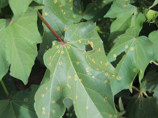
The top of a cotton leaf with rust colored spots surrounded by a yellow halo
Southwestern cotton rust caused by Puccinia cacabata
(Photo: J. Idowu, NMSU)
Southwestern cotton rust, caused by Puccinia cacabata, has been found on cotton plants in Southern New Mexico. This disease occurs sporadically in the Southwestern U.S. but has the potential to cause serious economic losses up to 50% under favorable environmental conditions. The disease has a complex lifecycle requiring two different host plants, cotton (Gossypium spp.) and grama grass (Bouteloua spp.), to complete a full disease cycle. During summer rains, the spores produced on grama grass germinate to produce airborne spores which are carried up to eight miles and cause initial infections in cotton. There is no repeating spore stage on cotton. All new infections on cotton are dependent upon spore showers from grama grass. The spores produced on cotton can only infect grama grass. Disease incidence is usually erratic in New Mexico and depends on summer rains, high humidity and an infected source of grama grass for inoculum.

The underside of a cotton leaf with orange colored spots cause by a rust fungus
Rust lesions on the underside of a cotton
leaf (Photo: NMSU - PDC)

A composite photo of cotton leaves with spots cause by rust
Rust lesions on the upper (left) and lower
(right) leaf surfaces (Photos: J. Idowu, NMSU,
left; and NMSU - PDC, right)


A composite photo of cotton leaves infected with rust. The photo on the left shows and early infection with small spots and the photo on the right shows a older infection with large dark brown spots.
Young cotton rust lesions (left) and older cotton rust lesions (right)
(Photos: NMSU - PDC)