Thursday, August 27, 2015

Featured Diagnosis: Bacterial Leaf Spot of Cucurbits

A pumpkin leaf with a large brown spot surrounded by a yellow halo
Bacterial leaf spot on pumpkin leaf. Note the yellow halo
surrounding the dark lesion.  (Photo: NMSU - PDC)
Bacterial Leaf Spot of Cucurbits – Bacterial leaf spot of cucurbits is caused by the bacterium, Xanthomonas campestris pv. cucurbitae. This disease causes sporadic losses in cucurbit crops grown in temperate climates. In New Mexico, the disease is not common, but can occur when warm, humid conditions are persistent. The disease attacks a number of different hosts including pumpkin, cucumber, gourds, and summer and winter squash.

Symptoms may appear on both the foliage and the fruit. On the foliage, the disease causes small somewhat round water-soaked lesions on the underside of the leaf. A yellow spot appears on the upper leaf surface. In a few days, the spots turn brown with a distinct yellow halo. The appearance on fruit is variable and depends on rind maturity and how much moisture is present. Initial lesions are typically small, slightly sunken, mostly round spots with a tan to beige center. As the spots enlarge (reaching up to 15 mm in diameter), they become noticeably sunken and the rind may cracks. 

A pumpkin with scabby, water soaked spots caused by a bacteria
Sunken bacterial leaf spot lesion on pumpkin
(Photo: J. French, NMSU-PDC).

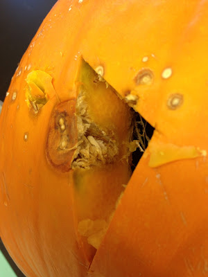
A pumpkin with a section removed to show the dark, watersoaked tissue  under the skin
Bacterial leaf spot lesion extends into the seed cavity
 (Photo: N. Goldberg, NMSU-PDC)

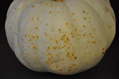
Small scabby lesions caused by bacteria on the outer skin of a white pumpkin
Bacterial leaf spot on a white pumpkin
(Photo: J. French, NMSU-PDC)


Wednesday, August 26, 2015

NMSU's Extension Service will host Plant Clinics at the Portales and Clovis Farmers' Markets

NMSU’s Extension Service will bring Plant Clinics to farmers’ markets in Portales and Clovis

New Mexico State University’s Extension Plant Sciences department will host a Plant Clinic at several farmers’ markets this summer and fall, to help educate the community about horticultural practices and pest management, including identification of insect, weed, and disease problems.

“We encourage anyone who is interested in gardening and has a question that they have not been able to get answered, to come out and visit with us,” said Natalie Goldberg, department head of NMSU’s Extension Plant Sciences. “If they have problem plants or pests that they would like to have identified, they are encouraged to bring us a sample of the plant or pest.”

The question-and-answer sessions will be hosted by four specialists: Carol Sutherland, entomologist; Leslie Beck, weed scientist, and Jason French and Goldberg, who are experts on plant diseases and disorders. In addition, the county Extension agent will be available to help answer questions.

“Some problems people bring to the clinic might be resolved on the spot, but others might need more background information to complete an answer, so we’ll make sure we have the client’s contact information and specific question written down so we can contact them later,” said Sutherland, who will have a collection of insects on display for additional questions.

Goldberg added they hope to answer most questions but if they can’t, they will either take back samples for further analysis or connect clients with other experts.

“Plant Clinics are a great way to interact with people not familiar with the great variety of information available through the Cooperative Extension Service,” Sutherland said.

Plant Clinics will be hosted at several Farmers' Markets around the state beginning in Portales (August 31 at 5 PM) and Clovis (September 1 at 5 PM).

Plant Clinics will also be held at the following farmers' markets:
September 12, Farmington, Farmington Farmers’ Market, 9AM
October 9, Lordsburg, Lordsburg Farmers’ Market, 4 PM
October 10, Mimbres, Mimbres Valley Harvest Festival, 10 AM
October 16, Belen, Belen Farmers’ Market, 4:30 PM
October 17, Albuquerque, Downtown Growers Market, 8:30 AM and Los Ranchos Growers Market, 8:00 AM


Image of advertising flyer for the Portales Plant Clinic
Flyer for the Farmers' Market Plant Clinic
in Portales, NM

Advertising flyer for Clovis Plant Clinic
Flyer for the Farmers' Market Plant Clinic
in Clovis, NM
A woman holding a large insect
Dr. Carol Sutherland identifies a beetle at a plant clinic in Alamogordo
(Photo: N. Goldberg, NMSU-PDC)

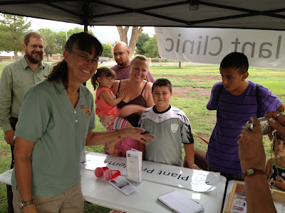
Image of people getting information at a plant clinic
Plant Clinic at the Las Cruces Farmers' Market
(Photo: J. French, NMSU-PDC)

Monday, August 24, 2015

Blossom End Rot

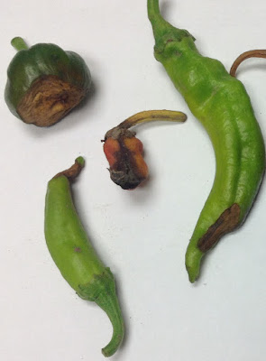
Image of pepper fruit with sunken, leathery brown spots at the blossom end of the fruit
Blossom end rot on peppers (Photo: N. Goldberg, NMSU-PDC)
Featured Diagnosis – Blossom end rot. Every year many home gardeners and commercial growers become concerned about a large tan to black spot on the bottom of fruit, especially on peppers and tomatoes. No fungi, bacteria, or any other living disease organisms are known to cause the condition, and it is not spread from one plant to another. This is “blossom-end rot,” a physiological disorder caused by a calcium deficiency in the developing fruit compounded by an imbalance in water and plant nutrients. It is especially problematic in the heat of the summer. As the weather warms and the plants begin to grow more rapidly, requirements for water and calcium increase. Because calcium is not a highly mobile element, a fluctuation in water availability—even for a short period—can result in a deficiency. It is at this time that fruit begins to show symptoms of blossom-end rot.

The disorder first appears as a brown discoloration on the blossom end of the fruit (the end opposite the stem). On chile fruit, the spot occasionally will be off to the side of the blossom end. The spot enlarges as the fruit matures and may eventually cover up to half the fruit. With age, the lesion tissue becomes sunken and leathery. Eventually, secondary fungi or bacteria may invade the tissue. Secondary invasion results in a black or watery appearance. Affected fruit ripens faster than unaffected fruit.


Image of pepper fruit with large tan spots on the blossom end of the fruit
Blossom end rot on peppers (Photo: J. French, NMSU - PDC)

Image of tomato fruit with large dark brown spots at the blossom end of the fruit
Blossom end rot on tomato fruit (Photo: NMSU-PDC)

Image of pepper fruit dark brown spots at the blossom end of the fruit
Blossom end rot on pepper fruit (Photo: J. French, NMSU-PDC)


Tuesday, August 18, 2015

Featured Diagnosis: Crown Gall (Agrobacterium tumefaciens)

Image of a large bacterial gall on a tree trunk
 Tree trunk infection by Agrobacterium tumefaciens. Resultant gall
nearly 2 feet across. (Photo N. Goldberg NMSU - PDC)
Crown gall, caused by the soilborne bacterium Agrobacterium tumefaciens, has a wide host range among woody and herbaceous plants. More than 600 plant species in over 90 plant families are susceptible. Some of the most common species affected in New Mexico include: apple, cottonwood, elm, grapes, juniper, pear, poplar, pecan, purple-leaf plum, pyracantha, roses, stone fruits, and willows. The bacterium stimulates host cells to divide and enlarge, causing tumor-like galls to develop. Galls on woody plants usually occur on roots and/or trunks at or just below ground level (the root crown) and at graft unions. The bacterium may become systemic in some host plants and cause galls on trunks, stems, branches and leaves above the root crown. Galls may also develop above the crown by pruning with infested cutting shears. On herbaceous plants, galls form on the roots and stems and, occasionally on leaves. Initially, galls are whitish, soft, and spongy. Later, the gall develops an irregular, rough, corky surface and a hard or woody interior. It eventually turns brown or black and may slough off the plant. Galls increase in size as the plant grows and may be less than an inch in diameter to several feet in diameter. The galls impede water and nutrient movement in the plant. Reduced transport of water and nutrients causes chlorosis, stunting, slow growth, and a general decline in plant health.


Large gall occurring at the graft union on a rose plant
Crown gall infection at the graft union on rose.
(Photo N. Goldberg NMSU-PDC)

Very large gall at the root flare of a large tree
Root flare infection by crown gall. (Photo: N. Goldberg NMSU-PDC)

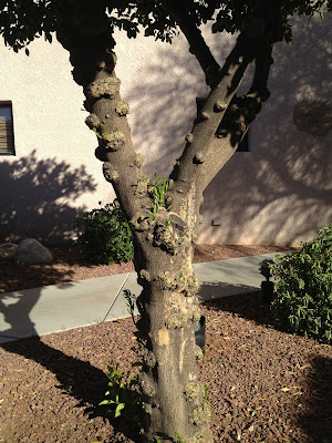
Golf ball to baseball sized galls occurring all over the trunk of a tree
Severe crown gall infection on the trunk and
branches of a lime tree.
 (Photo: N. Goldberg NMSU -PDC)

Quarter sized galls on the roots of a peach tree
Large galls on peach tree roots caused by
Agrobacterium tumefaciens
(Photo: J. FrenchNMSU - PDC)

Tuesday, August 11, 2015

Featured Diagnosis: Dodder (Cuscuta spp.)

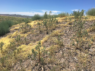
A landscape with yellow mats of a parasitic plant covering the plants and ground
Large mats of dodder parasitizing weeds on a disturbed
road side site. (Photo J. French NMSU-PDC) 
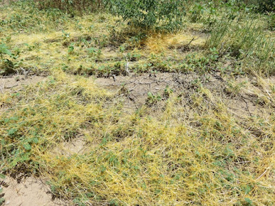
Environmental conditions across the state are perfect for the parasitic weed, dodder, to germinate and grow. This parasite does not have leaves and cannot produce chlorophyll. As such, it grows on other plants, using them for water, nutrients, and carbohydrates. It is common along road sides, but can also grow on crop and landscape plants. In the spring, dodder seeds germinate near the soil surface and send up slender, thread-like twining stems varying in color from pale green to yellow or orange and without any cotyledons (seed leaves). The slender, leafless, thread-like stem sways or rotates slowly until it touches the stem or leaf of another plant and begins to wind around it. On a host plant, the dodder stem will immediately form small appendages called haustoria (tiny sucker-like roots), which penetrate the stems or leaves so that dodder can extract its necessary growth requirements. Soon after attaching to a host plant, the lower end of the dodder withers and breaks its connection with the ground, while the upper part of the stem grows rapidly, often forming dense stringy masses. However, if the dodder seedlings are unable to contact a susceptible host plant soon after germination, they will not survive. The damage of dodder to the host plant varies from moderate to severe depending on the growth of the host plant and on the number of haustoria attachments to the host plant. Dodder infestations reduce crop yield and increase harvesting costs for crops like alfalfa. 

For more information please visit Dodder (Cuscuta spp.)


A close-up of a landscape with yellow mats of a parasitic plant covering the plants and ground
Dodder in a native landscape setting. (Photo L. Beck NMSU-PDC)

Microscopic view of a parasitic plant penetrating the stem of a host plant
Dodder haustoria (tiny sucker-like roots) penetrating the stem of a host plant.
The parasite uses these projections to extract the necessary nutrients
to 
survive. 
(Photo J. French NMSU-PDC)

A composite photo showing the flowers of the parasitic plant dodder. The photo insert shows a close-up of the white flowers.
Small clusters of dodder flowers forming along the stem. Each flower will
produce a small seed pod with 2 to 4 seeds. (Photo J. French NMSU-PDC)

Wednesday, August 5, 2015

High Humidity Encourages Turf Diseases

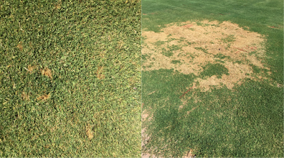
A composite photo of turfgrass with brown dead areas caused by the fungus, Bipolaris. The photo on the left shows small brown areas when the disease was in the early stages and the photo on the right shows the same turfgrass area 10 days later with a very large brown area.
Turfgrass infected by Bipolaris sp. - these photos are the same turfgrass
area photographed 10 days apart (Photos: NMSU - PDC)
Summer rain is a welcome occurrence in the desert, however it can also create a favorable environment for diseases. Recently, conditions have been especially favorable for a few common turfgrass diseases. Leaf spot, melting out and brown patch, have been identified in a number of samples submitted to the plant clinic over the past several weeks. These diseases are caused by common soil-borne fungi that are favored by wet, humid conditions. They can be especially severe when heavy rains follow periods of drought or dry conditions. Leaf spot and melting-out diseases are caused by a group of pathogens that used to be grouped together in the fungal genus Helminthosporium. Two fungi in this group, Bipolaris sp. and Curvularia sp., have been isolated from turfgrass samples in New Mexico. Brown patch is caused by Rhizoctonia solani. Similar environmental conditions favor these diseases and it is not uncommon to find more than one of these pathogens in the same sample. When
environmental conditions are highly favorable for disease development, these diseases can spread rapidly resulting in large areas of blighted turf.


A composite microscope photograph of spores of the Curvularia fungus
Curvularia spores
(Photo: NMSU - PDC)
A blade of turfgrass with small dark brown spots with a tan center
Leaf spots
caused by
Bipolaris
(Photo: NMSU-PDC)
A composite microscope photograph of spores of the Bipolaris fungus
Bipolaris spores
(Photo: NMSU - PDC)


Turfgrass with a large patch of brown (dead) grass
Brown patch caused by Rhizoctonia solani
(Photo: NMSU - PDC)
A microscope photo showing the characteristic hyphae (fungal strands) with right angle branching of Rhizoctonia solani
Characteristic hyphae of Rhizoctonia
solani
(note the right angle branching)
(Photo: NMSU - PDC)

Tuesday, August 4, 2015

Featured Diagnosis: Honey locust Agrilus (Agrilus difficilis)

A composite photo of the insect, Agrilus difficilis. The photo on the left shows the top of the insect and the photo on the right shows the underside of the insect.
Upper and lower surfaces of adult Honey Locust Agrilus.
Actual size, ~ 3/8” long Photo:
J. Shaughney NMSU Arthropod Collection
    
Agrilus difficilis, the ‘honey locust Agrilus’ and a close relative of ‘Emerald ash borer’ was identified in Central New Mexico infesting honey locust (Gleditsia triacanthos). The tree canopy was leafless and apparently lifeless down to some green sucker growth near the ground. Upon closer examination there were D-shaped emergence holes in the bark, peeling bark with multiple larval feeding trails on its inner surface and small, very thin, blackish beetles that died in their attempts to escape their host. All of these beetle-related observations are similar to those caused by ‘emerald ash borer,’ an exotic, invasive ash-tree killer not known to occur yet in New Mexico. Dr. Carol Sutherland presumptively identified the insect as  Agrilus difficilis, the ‘honey locust Agrilus’ and this was later confirmed by Dr. Zablotny of the United States Department of Agriculture. 

Image of D-shaped emergence holes on a tree trunk caused by Agrilus difficilis
Emergence hole (D-shaped) made by an adult
 leaving the tree. 
R. Husted, Sandoval Co. MG. 


Photo of damage under the bark. There are adult insects stuck in the emergence holes.
Inside of honey locust bark – severe vascular damage from
larval stage. The black objects are adult A. difficilis that died
 before it could chew its way through the bark.
Photo C. Sutherland NMSU-PDC
    
Image of the woman standing by a nearly dead tree
This infested honey locust died
from the top down note the green
 sucker growth at the bottom.
Photo: R. Husted, Sandoval Co. MG.


Photo showing two white larvae with dark heads and two sharp spikes at the rear
A. difficilis larvae extracted from honey locust. The two little
dark brown ‘splinters’ on the rear are characteristic
of certain Agrilus species. Photo C. Sutherland NMSU-PDC