Thursday, May 11, 2017

Featured Diagnosis: Heuchera Rust

small brown fungus pustules on the underside of a leaf
Rust pustules on the underside of the leaf
(Photo: J. French, NMSU-PDC)
Rust on coral bells is caused by Puccinia heucherae, which only infects members of the genera Heuchera and Saxifraga. A wide range of plants can be infected by rust fungi, but individual rust fungi have a very limited host range. The most common symptom of rust infection is the production of powdery pustules on the leaves, stems, twigs, flowers and fruit of susceptible plants. Pustules are most common on the lower leaf surface and stem. Pustules may be bright yellow, orange, orange-red, reddish brown, chocolate brown, or black in color. These spots enlarge and may coalesce as the infection builds. Rusted leaves often turn yellow, die, and drop prematurely. Rust fungi infect only the plant’s aboveground parts, and while rust fungi generally do not directly kill their host plants, severe infections ultimately may lead to death by other factors (winter-kill or other diseases). Spores are moved short distances by wind, insects, rain, and animals. Water on the plant surface (aka leaf wetness) is required for spore germination and infection. After the plant is infected, leaf wetness is not needed for continued disease development. Disease builds quickly during periods of high humidity. 


small yellow spots on the upper leaf surface
Rust on the upper leaf surface of
coral bells (Photo: Jason French)


A microscopic photograph of rust spores
Teliospores of Puccinia sp. (Photo: Jason French, NMSU)

Friday, December 2, 2016

Stem and Bulb Nematode Discovered in New Mexico

composite photo showing garlic bulbs with damage from stem and bulb nematode
Figure 1. Garlic plants exhibiting severe symptoms of stem
and bulb nematode (Photo: J. M. French, NMSU-PDC)
Stem and bulb nematode, Ditylenchus dipaci, discovered on Garlic in the Rio Grande Valley of New Mexico - Ditylenchus dipsaci is a serious temperate-climate nematode pest of over 500 plant species in over 40 plant families, including alfalfa, onions, and garlic.  Among plant-parasitic nematodes, it ranks fifth in economic importance and is a quarantine pest of international concern. In 2015, New Mexico harvested 190,000 acres of alfalfa hay with a value of over $188M and is the eighth largest producer of onions in the U.S. cultivating 5,200 acres in that year with a crop value of $91M. These two crops rank number 1 and number 3, respectively, for highest grossing crops in NM in 2015. New Mexico also has a vibrant small farm garlic industry that supplies local markets with fresh garlic and seed for producers.  New Mexico has never reported an infestation of D. dipsaci and its presence could have serious ramifications for NM’s alfalfa, onion, and garlic producers. 

In May of 2015 garlic plants (Allium sativum) from a home garden in were submitted to the Plant Diagnostic Clinic at NMSU.  The grower reported poor growth in ~30% of 1,200 plants originating from seed purchased in southern New Mexico.  Early symptoms included chlorosis, wilting, and poor root and bulb development.  As the disease progressed, roots turned brown, were easily separated from the bulb, and older foliage had collapsed and turned straw colored (Figure 1).  Microscopic examination showed large numbers of nematodes present in symptomatic garlic plants. Based morphological characteristics of the nematode and the DNA sequence of the ITS-1 region, the nematodes were identified as D. dipsaci.

This discovery is highly significant as this nematode has the potential to cause significant economic losses on agriculturally important hosts grown in the state and in the region. The longevity of this pest in the soil and international trade issues are major concerns for producers. Based on information from the grower it appears that this is an isolated introduction from infested plant material which can potentially be contained as the presence of D. dipsaci was discovered before any material was harvested or distributed. Monitoring of production areas in the state will be performed to determine if this was an isolated and contained introduction or if this important pest has become established in NM.
 
microscopic photograph of a stem and bulb nematode from a garlic plant
Stem and bulb nematode isolated from garlic
(photo: J. Beacham, NMSU)

Friday, October 7, 2016

Plant Clinics in Albuquerque this Weekend

Specialist's from the NMSU - Plant Diagnostic Clinic will be holding plant clinics this weekend in the Albuquerque area. We will be at the Downtown Growers' Market on Saturday morning from 8 AM- 11:30 AM and at the Local Food Festival at the Gutierrez - Hubbell House on Sunday from 10 AM - 2 PM. Please come and visit us! If you have a problem plant or pest, bring it along, we'll do our best to identify the problem and provide information on management options! (The photo below is from the plant clinic at the Local Food Festival two years ago). Hope to see you there!

Photo of NMSU employees at the Local Food and Field Festival in Albuquerque, NM in 2014
NMSU Faculty at the Local Food Festival in Albuquerque in 2014

Wednesday, September 21, 2016

Tomato Spotted Wilt Virus Strikes Again

A chile pepper plant with leaf deformity, spotting and yellowing caused by a virus
Chile plant exhibiting symptoms of Tomato
Spotted Wilt Virus (Photo: NMSU - PDC)
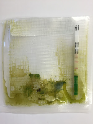
We've highlighted this disease before, but it's such an interesting disease that we decided to do it again! This chile plant was submitted for disease analysis exhibiting classic symptoms of Tomato Spotted Wilt Virus (TSWV). Symptoms of TSWV can be quite variable, but some of the more common symptoms include leaf deformity (especially of the new growth), terminal necrosis, leaf drooping (wilt-like appearance), stunting, mottling, and spotting (necrotic and chlorotic). In this particular case, several leaves on the plant displayed characteristic chlorotic spots with concentric rings (see images below).  Fruit symptoms can also be quite striking. Fruit symptoms include: chlorotic concentric ringspots, raised bumps, uneven ripening and deformity. Plants infected early in the season may produce little or no fruit. In this case, the plant produced only one small, deformed fruit which ripened prematurely. Most viral diseases are confirmed using some form of DNA analysis. In this case, there is an antibody-antigen test that can detect the virus in just a few minutes. A small amount of plant tissue is ground in buffer and then a strip embedded with antibodies to the virus is placed in the sap solution. If the virus is present, two purple lines appear on the strip (if only one line appears, it indicates that the test worked, but the virus was not present). Below is a photo of the antibody-antigen test for this plant.


The terminal branches of a chile plant with leaf curling and deformity caused by a virus
Terminal leaf deformity caused by Tomato Spotted
Wilt Virus (Photo: NMSU-PDC)

A composite photo of chile pepper leaves with chlorotic concentric ringspots caused by a virus
Chile pepper leaves exhibiting chlorotic concentric ringspots characteristic
of TSWV (Photo: NMSU-PDC)

A plastic sample bag with a crushed up chile peper leaf and an indicator stick showing the leaf sample is infected with a virus
Antibody-antigen test results for Tomato Spotted Wilt Virus -
two purple lines indicated that the tissue is infected
with Tomato Spotted Wilt Virus (Photo: NMSU-PDC)

Tuesday, September 13, 2016

Tubakia Leaf Spot

Tubakia Leaf Spot

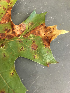
Oak leaves with irregular brown spots caused by a fungus
Tubakia leaf lesion on Oak.
Tubakia sp. a fungal pathogen that causes a late season leaf spot and twig canker. All species of oak appear to be susceptible to the disease but those in the red oak group ( black, red & pin oak) appear to be most susceptible.

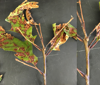
Tubakia leaf spot tends to develop in the late summer to early fall and begins as small reddish-brown foliar spots. As the disease progresses the leaf spots enlarge and may coalesce to form large blighted areas. Spotting may occurs on or near leaf veins causing death of the vein and collapse of leaf tissue beyond that point. Premature leaf drop and small twig cankers can form in severe cases.

Tubakia sp. overwinter in twig cankers and in infected leaf tissue on or around the tree. Spores are easily spread by wind and rain splash to uninfected tissue in the spring and early summer months. In most cases, Tubakia leaf spot is a cosmetic disease. In general, when leaf spots are sporadic, the ultimate damage to the plant is relatively insignificant. When leaf spots occur year after year on the same tree, cumulative damage may result in dieback and decline.

Management of Tubakia begins with good sanitation practices such as raking and destroying fallen leaves. This reduces the initial inoculum for the following year. Selectively pruning branches to increase air circulation within the tree canopy will further reduce the incidence and severity of the disease. Branches exhibiting cankers or dieback should also be removed from the tree. This will improve the appearance of the tree as well as reduce overwintering sites for the pathogen. Lastly ensuring that the tree is properly cared for (irrigation and fertility programs) will reduce plant stress and decrease susceptibility to the disease.

Close up of irregular brown spots on an oak leaf caused by a fungus
Tubakia sp. causing leaf vein death and
collapse of leaf tissue beyond that point.
Oaks leave dying and turning light brown due to fungus infection
 Advanced stages of Tubakia leaf spot note coalescing
lesions, twig cankers and premature defoliation.


Microscopic photograph of fungus spores
The conidia (dark, ovoid structures pictured here) are produced
by the fungus and serve as inoculum for new infections.


Thursday, June 16, 2016

Tomato Psyllids

picture of tomato psyllid adults and nymphs
Tomato psyllid adults and nymphs (Photo: NMSU-PDC)
Featured Diagnosis: Tomato (Potato) Psyllids - Hemiptera, Psyllidae, Bactericera cockerelli.


These tiny insects will attack a wide range of hosts but prefer solanaceous plants like tomato, potato, peppers, and eggplant. Published reports indicate that yellow pear tomato is especially susceptible to tomato (potato) psyllids. The infestation pictured here began on a yellow pear tomato and quickly spread to several other tomato varieties. Psyllid eggs are laid on stalks on the under side of the leaves. Nymphs hatch from the eggs and are flattened, oval and yellowish green to orange with red eyes and three pairs of legs. They develop through five instars before becoming adults. Adult psyllids are ~3mm long have white to yellowish markings and clear wings. Psyllids feed most often on the underside of the leaves. While feeding, the insect injects a toxin into the plant. The toxin causes a variety of symptoms which collectively are commonly referred to as “psyllid yellows.” These symptoms include stunting, yellowing, and curling of the leaves. Plants damaged by psyllids may produce little or no fruit. Fruit that is produced is often small, even in large-fruited varieties, and of poor quality. Small seedlings or transplants may be killed by the insect. 


A composite photo of tomato plants infested with small insects called psyllids
Tomato psyllids on leaves and stems of tomato plants (Photos: NMSU-PDC)


tomato plant in a pot with psyllid insects feeding on it
Tomato psyllid infested tomato plant (Photo: NMSU-PDC)


Wednesday, June 15, 2016

Tomato Spotted Wilt Virus on Dahlia

Dahlia plant with yellow flecking on the leaves caused by a virus
Tomato Spotted Wilt Virus on dahlia - note
the yellow flecking near the tip of the leaf
(Photo: NMSU-PDC)
Featured Diagnosis: Tomato Spotted Wilt Virus is a common virus with the ability to cause disease on a wide range of hosts. In New Mexico, it is often found on peppers and tomatoes. Recently, the virus was identified on dahlia. Dahlia is a known host for this pathogen, but this is the first report of the disease on dahlia in New Mexico. The symptoms on the dahlia samples submitted to the NMSU Plant Diagnostic Clinic were subtle; a yellow flecking on some of the leaves.